Friday, December 30, 2011

Micro-rock-pooling in Winter.


It's too cold in winter to spend a lot of time paddling around in rock pools but you can always take a few samples of seaweed home on a jar of seawater and have a look at the smaller inhabitants under the microscope. These two, each about a millimetre long, were in a  sample of Corallina officinalis seaweed. The upper specimen is an unusually bristly acarine mite, found clambering through the seaweed fronds. You can see more acarine mites by clicking here.

This is a minute flatworm, with two very simple eyes, found gliding over the surface of the seaweed, propelled by thousands of cilia that are only visible at high magnification under the microscope. You can see another marine flatworm, in more detail and with a movie of the cilia in action, by clicking here.

Friday, December 23, 2011

Sowing Wild Oats


Some seeds need to be sown while others - like wild oat Avena fatua - sow themselves. This is a wild oat fruit (or, to be botanically accurate a caryopsis) in the dry state. It's equipped with a long awn (which is extension of the floret in which  the fruit formed and in which it is shed), that's bent at a right angle about a quarter of the way along it's length.


When the caryopsis falls to the ground and gets wet - from a passing shower of rain, for example, that bent awn straightens, then bends again as it dries out. The picture above shows the same fruit, but now it's been moistened and the awn has straightened. As the awn bends and straightens it also rotates, because the awn is constructed from a helix of fibres that twist and generate torsion as they dry (see below).


The outer coat of the floret containing the caryopsis is equipped with this arrowhead of stiff hairs at the tip ....


.....which readily catch in fur and feathers and help disperse the seed, but also anchor it in crevices in the soil when it falls to earth.


There is also a beard of stiff hairs running up the groove in the caryopsis. As the awn rotates .....


.... with the expansion and contraction of this helical tube of spiral fibres that it's constructed from, it levers the caryopsis further into soil crevices. Those stiff hairs on the caryopsis help to anchor it in the soil, ratcheting it in ever deeper until it's in a  moist enough position to germinate and put down roots. 

This is a seed that sows itself.

The video below shows a group of wild oat caryopses writhing as their awns dry out and begin to rotate.


Sunday, November 27, 2011

Breathing Space


This is a thin section of the lower stem of water milfoil Myriophyllum sp. , stained with the fluorochrome calcofluor which binds to the cellulose of the cell walls and is fluorescing brightly in ultraviolet light. Marsh plants tend to be rooted in anaerobic mud and so have air channels (aerenchyma) that conduct oxygen down to the roots. 


Working from the outside inwards in this section, there is a well defined single outer layer of very small cells forming the epidermis, then inside that lies the stem cortex with 17 air channels arranged around the central stele, which contains the phloem (brightest flourescence) and the xylem.


The stem and leaves of water milfoil. The small white structures in the leaf axils are the stigmas of the female flowers.

Thursday, November 10, 2011

Plant Cuticles


The surface of plants (with a few exceptions, such as those that live submerged under water) is covered with a tough, transparent, waxy layer called the cuticle, composed of cutin secreted by the layer of epidermal cells that it covers. The best way to see the cuticle is to snap the leaf of a drought-adapted succulent plant like Crassula ovata and pull one part of the leaf against the other, peeling away the cuticle, which covers the above ground parts like a wrapping of cling-film.


These  cuticular peels, often with a single layer of epidermal cells attached, can be mounted on a microscope slide .....


..... and viewed to reveal the pattern of cells and the .....


...... stomata, which allow carbon dioxide to enter for photosynthesis. The two images above are of cuticular peels of spiderwort Tradescantia virginiana.


In this vertical section of a leaf, stained with fluorescent dyes, the cuticle appears as the bright yellow layer on top of the epidermal cells. In the centre you can see a single stoma in vertical section, with closed guard cells and its adjacent subsidiary cells, with a sub-stomatal cavity below leading to the loosely packed mesophyll cells, where photosynthesis takes place. The cuticle has a dual function - keeping water in and keeping the leaf surface dry. 


In plants adapted to arid conditions (xerophytes), like this Aloe variegata  , the cuticle is visible as an extremely thick transparent layer that allows very little water to escape from the leaf.


In plants that are subjected to frequent rainfall, like this nasturtium Tropaeolum majus leaf, fine grooves in the cuticle surface trap air below the water droplets, which then round-up under their own surface tension and simply roll off the leaf when it shakes in the wind. This is vital, as a wet leaf surface blocks stomata and prevents carbon dioxide from entering, slowing down photosynthesis. As the water rolls off the leaf it carries away dirt and dust, so the leaf cuticle is effectively a self-cleaning surface - a phenomenon known as the 'lotus effect', referring to the extremely hydrophobic self-cleaning leaves of sacred lotus. The silvery area under the central water droplet in the image above is caused by minute air bubbles, trapped between the water and the leaf surface.


The cuticle can also have a secondary defensive role, as seen in this painfully prickly leaf of the hedgehog holly Ilex aquifolium 'ferox', which is covered in cuticular spines. In general, leaves of evergreens, that survive for several years before they are shed, tend to have thick cuticles that protect the leaf against herbivore attack throughout their extended life.


Thursday, November 3, 2011

Springtails


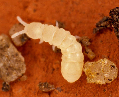
Every time I lift the lid of our garden compost bin scores of these tiny insects, each smaller than the diameter of the head of a pin, leap around in all directions. They are members of the ancient insect order known as the Collembola - commonly called springtails - and feed on decaying vegetation. The darkness, warmth and humidity of the compost bin suits them perfectly. I think this species might be Folsomia candida, which is very common in gardens.


Most of the time they move slowly on those stumpy legs but when they are alarmed they hurl themselves into the air using an organ called a furcula under their tail .....

.... which you can see in this specimen. You could liken its action to a kind of exceptionally energetic pole-vaulting. The tip of the furcula is held in place by a clip-like structure called a retinaculum, but when the muscles in the furcula contract the clip suddenly releases its grip and the furcula flicks downwards and backwards, hurling the animal upwards and forwards.

You can find pictures of another springtail species here and a fine set of photographs for ID purposes here.

Monday, October 31, 2011

Was Sid, the Mystery Microscopist, a Scotsman?


When I was exploring the old microscope slides belonging to the mysterious 'Sid' that I mentioned in my previous post I came across one that was clearly home-made and was labelled as porphyritic olivine basalt from the Lion's Haunch, Arthur's Seat, Edinburgh. The hill is the plug from the mouth of a long extinct volcano, exposed after softer sedimentary rocks were eroded from around the igneous core.  Polarised light microscopy reveals the crystalline inclusions very nicely.

Similar rock samples were collected on the Apollo 15 mission to the moon in 1971

Arthur's Seat is the very conspicuous hill - some might call it a mountain - almost in the centre of Edinburgh. From the right angle it looks like a resting lion and this polished thin section of igneous rock came from the 'Lion's Haunch'. Of couse, there's no way of telling whether Sid, who was apparently a chemist, prepared this specimen himself or swapped it with a geologist friend - microscope slide exchange clubs were once common - but he clearly had an interest in Scottish geology, so may have been a Scotsman......


Friday, September 30, 2011

Mystery Microscopist

Many years ago, when I was a student, I was given this box of old prepared microscope slides. I can't remember who the donor was but they've been at the back of a cupboard for years and only emerged when I was having a clear-out, a couple of months ago.


They date from around the time of the First World War. Some were commecially prepared by the firm of Watson & Son, of 313, High Holborn, London - labelled with beautiful handwriting, in mounts that were so well ringed with shellac that they have remained in perfect condition for almost a century.

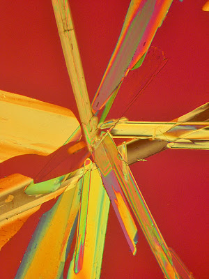
Others were prepared by the original owner, who I'm guessing must have been a chemist because many are cystalised mounts of chemical compounds, intended to be viewed with polarised light, made from substances that would only have been available to a professional chemist - possibly a plant biochemist because quite a number of the crystals are naturally-occuring plant compounds that he might have extracted and purified himself. One contains the only clue to the identity of the mystery microscopist, because he has written his name in chemical crystals on the slide - 'SID'.

Sid would have looked at these specimens with a rather primitive instrument called a Fox Polariscope - so Sid probably wouldn't have seen the images in quite the same vibrant colours that you can see here, achieved with a modern polarising microscope. The specimen above is strychnine...


... this is floridzin, an alkaloid from apple roots...

...as is this one, too.

This is mercuric cyanide

Salignin...

Coumarin, the compound responsibe for the scent of new-mown hay...

Salicin, extracted from willow bark and the precursor of salicylic acid ,better known as aspirin...

... and ammonium bitartrate


Friday, September 23, 2011

Hooked on Hops

Hops Humulus lupulus have an impressive ability to climb supports - either up other plants or, in the case of cultivated hops, up poles in hop gardens. Charles Darwin devoted a lot of time to studying the way in which their shoot tips rotate as they grow (by the process of circumnutation), seeking out objects to coil around (you can read more about his experiments here). There's more to hops' climbing ability than circumnutation and rapid growth, however - their stems are clothed in very distinctive epidermal hairs (trichomes) that act as grappling hooks, securing their grip on supporting structures.

 
The hop trichomes that are adapted for climbing have a very distinctive anvil shape - you can see them here, at low magnification, on either side of a hop leaf petiole.

At higher magnification the anvil shape is very distinctive, something noted ....

.... by the botanist Anton Kerner von Marilaun in his Natural History of Plants (1895).

Hops have been cultivated for centuries, primarily for the resins produced by their epidermal glands, mainly at the base of the bracts in the female flowers but also on other parts of the plant, including the underside of the leaf. In the photograph above you can see the minute gold drops of resin on the lower surface of a hop leaf. The resins are converted to bitter isohumulones during the brewing process, adding a distinctive flavour to beer.


Monday, August 22, 2011

Bacterial Root Nodules

These are bacterial root nodules on the root of runner bean Phaseolus coccineus. Each nodule contains a population of Rhizobium bacteria that are capable of converting atmospheric nitrogen into soluble forms of nitrogen that the plant can use for growth - which is what makes this symbiotic association between plant and bacterium so valuable for agriculture. In annual legume crops, once the bean crop has been harvested the root nodules decay and release nitrogen in the soil, where it can give a yield boost to following non-legume crops in the crop rotation - like wheat, for example.


In this image one of the nodules has been cut in transverse section and stained with the fluorochromes calcofluor and auramine O. The plant root, with its xylem vessels visible, is at the top. The bacteria filling the root nodule, encased in blue-stained plant cells, are stained yellow. The Rhizobium bacteria in the soil penetrate through a root hair, trigger proliferation of the host plant root cells to form a nodule and multiply within. Healthy root nodules are pink when you cut them open due to the presence of leghaemoglobin which, like haemoglobin in mammalian blood, absorbs oxygen. This is important because oxygen would otherwise inhibit the enzymes in the nodule that 'fix' nitrogen into soluble forms. Bacterial nodules that are not pink when you cut them open are likely to be parasitic on the host plant, rather than symbiotic.

This is the difference that nodulation makes. The plant on the right has effective nodules, the one on the left doesn't. The interaction between plant and bacterial strain is complex; for any give crop cultivar, different bacteral strains will show varying degrees of effectiveness in boosting crop yield and different crop varieties perform best with different bacterial strains. Deliberately inoculating seeds with effective Rhizobium strains can produce significant yield benefits, although there is no guarantee that any particular inoculum will persist in a soil type or location where it's not a naturally-occurring strain amongst the existing soil microbial community.


Wednesday, August 10, 2011

Out of Sight,Out of Mind...

Roots are the most neglected parts of plants, perhaps because they are out of sight and - superficially at least - lack the intrinsic aesthetic beauty of the above-ground parts. For most (although not all) plants they are vital structures and - when you look really closely - they have an intricate beauty of their own.

Root tips are sensitive gravity detectors, ensuring that the root always grows downwards into the soil. This root was held in the horizontal plane for less than an hour before it redirected its growth downwards. Behind the root tip you can see the point where the root hairs develop, with newly initiated root hairs just visible nearest the root tip but becoming longer as you move away from it. Further back still the root hairs die away continually and each has a life span of just a day or two, but they are continually replaced as the root penetrates further into the soil. The passage of the root through the soil is assisted by lubricating mucilage produced by the root tip, whose surface cells slough off. The mucilage also supports a bacterial microflora that helps the root acquire nutrients and may provide some protection from disease-producing organisms.

This is a root tip sectioned vertically and stained with a  fluorescent dye called DAPI. If you click on the image to enlarge it the details will be a little clearer. The brightly fluoresescing dots are the nuclei, one per cell, and you can see the files of cells produced by sequential cell division followed by cell elongation, which pushes the root ever-further into the soil.

This is a root in transverse section, further back from the tip than the previous image, in the middle of the root hair zone. It has been stained with a fluorescent dye called calcofluor, which makes the cellulose cell walls fluoresce blue in ultraviolet light. From the outside inwards, you can see the long root hairs, each a single cell that arises from the root epidermis (surface layer of cells). Next inwards lies the root cortex, which constitutes the vast bulk of the cells, then in the centre you can see the stele - the cylinder of vascular tissue that transports water upwards to the rest of the plant and carries sugars and amino acides downwards to support the continued growth of the root.

The arrangement of the various cells and structures is more clearly visible here, at higher magnification. The large circles in the stele, top left, are xylem vessels that conduct water away from the root.

The root hairs, which are in intimate contact with the soil particles, absorb water and soluble minerals that are transported through the root cortex, both from cell-to-cell within cell cytoplasm (the symplastic route) and through cell walls and the spaces between cells (the apoplastic route), to the stele in the centre of the root.


Once the water reaches the stele it encounters a single layer of cells called the endodermis, that sheaths the stele. The walls of the endodermal cells contain a substance called suberin which renders them impermeable, so water that arrived via the apoplastic route is forced into and through the cytoplasm of these cells, where dissolved minerals are selectively removed. You can see the suberin deposits, known as the Casparian strip, as the orange staining in the single ring of cells that lies between the blue and the yellow cells in the section of a stele above. Some water also passes unimpeded through specialised passage cells in the endodermis - if you follow the ring of cells with the orange stained Casparian strip in their cell walls around the stele in the image above, you'll notice a few passage cells with no orange-stained suberin deposit in their walls.
Almost all the water taken up and transmitted via both routes, via the cytoplasm of the endodemis cells or via their passage cells,  then enters the dead xylem cells that carry it aloft in the water column that is drawn upwards by transpiration from the leaves.

When gardeners buy plants in garden centres there's a great temptation to simply dig a hole and plant them, without teasing out the pot-bound roots or cultivating the soil around the planting hole, but a little tender, loving care for root systems pays great dividends: the vigour of the plant above the soil depends on the health of the roots, hidden below the surface.

Saturday, June 18, 2011

Banana Stellate Parenchyma

These beautiful cells come from the midrib of a banana leaf. Each is shaped like a 6- or 7-armed star, with its arms joined to the arms of surrounding cells, forming a lattice of cells. This form of tissue is known as stellate parenchyma and you can find another example here. The image was produced using polarised light and the brightly coloured birefringent objects inside the cells are calcium oxalate crystals inside the cell vacuole. You can see further examples of calcium oxalate crystals, including a video of their Brownian motion inside a cell, if you click here.


To find these cells you need to look inside the midrib of a banana (Musa sp.) leaf .....


by cutting transversely across the midrib, which reveals this internal pattern of strenthening tissue filled with very delicate, transverse plates of glassy cells ....

... then dissect out one of these plates of cells and mount it on a microscope slide.