Friday, December 24, 2010

Nature's Stained Glass Windows


Looking like a stained glass window, this is the remains of the seed pod of one of last summer's garden flowers, Lunaria annua (commonly known as honesty, because the dry seed pods resemble silver coins), magnified around two hundred times and viewed using polarised light.


When honesty seed pods ripen they are flattened and composed of three components. Imagine three large 'coins' joined to each other all around their rims, with the central 'coin' attached to the plant via a stalk. Swelling seeds are attached to the rim of both faces of the central 'coin' via their own slender stalks, visible when sunlight shines through the whole structure.

When the pod dries out and ripens the tensions in the drying, contracting cells of the walls of the 'coins' tear them apart around their rims, so the two outer 'coins' detach and flutter away in the breeze, followed by the winged seeds, leaving the central 'coin' attached to the dead plant and surviving deep into winter.

These are the cells of that central, surviving 'coin' magnified about one hundred times and using ordinary white light....


... and these are the dazzling interference colours generated when polarised light is used.


At two hundred times magnification it's clear that the 'coin' is formed from two layers of cells, orientated at different angles, so that the tensions they develop when they dry will twist and distort the 'coin' and help to rip apart the sutures with the outer 'coins'.


 At four hundred times magnification you can clearly see the pores through the thick cell walls which were the plasmodesmata - the channels of communication between the cytoplasmic contents of one cell and the next, while the whole structure was still alive and the seeds were still developing.




The dry, dead cells form intricate patterns...


... whose colours change as they are rotated in the plane of the polarised light.

Saturday, December 18, 2010

The Colour Purple

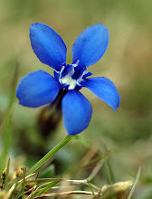
This rather beautiful flower is Tibouchina urvilleana and the purple of its petals is due to the presence of anthocyanin pigments, which are dissolved in the cytoplasm of the petal cells.


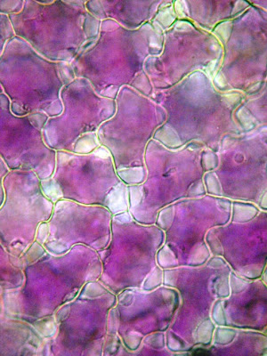
If you magnify the petal surface about 200 times you can see the way in which the petal cells fit together, like pieces of a jigsaw puzzle. This piece of petal is mounted in water but if it's transferred to a concentrated solution of sugar ...


  ... the water in the cell begins to flow out through the semi-permeable cell membrane by the process of osmosis, with water travelling out from the less concentrated solution in the cell to the more concentrated solution surrounding it. Within a few minutes spaces become visible between the cytoplasm and the cell wall, where the cytoplasm shrinks ....


... and within a few more minutes the cytoplasmic contents of the cell have shrunk even further, so the purple anthocyanin pigment becomes even more concentrated in the remaining cytoplasm.


  While I was looking at the petals I noticed something unusual around their edge  - a fringe of microscopic hairs, invisible to the naked eye.


 Each hair is tipped with a  glandular head.


... that looks as through it may contain oils.

What are these hairs for? Secreting aromatic compounds that attract insect pollinators, perhaps?

Friday, December 3, 2010

Sticky Jack

This is a cross section of the stem of the plant commonly known as goosegrass or sticky Jack and more scientifically as Galium aparine. Sticky Jack is a very common weed that scrambles up through other vegetation using its covering of hooked hairs on the stem and leaves and which sticks to clothing with these when kids throw handfuls of the stuff at each other.
This image was produced using fluorescence microscopy, staining the cells with compounds that bind to the cell walls and fluoresce. The blue cells have walls made of cellulose and their blue fluorescence is due to the calcofluor that they've been stained with, which fluoresces blue in ultraviolet light. Calcofluor has been used as a 'blue whitener' in washing powders - it binds to the cellulose in cotton fabrics and fluoresces faintly blue in the UV component of sunlight. The yellow staining is due to another fluorescent dye (fluorochrome) called auramine O, which binds to cutin in the outer cuticle of the plant, and to dead, lignified cell walls that give the stem its strength - and it fluoresces yellow. The cuticle in this cross section is the thin yellow line covering the outer surface of the section. The yellow circle in the centre is composed of dead, lignified cells - not particularly well developed in goosegrass because it scrambles over surrounding vegetation rather then investing resources in producing a stout lignified stem of its own.